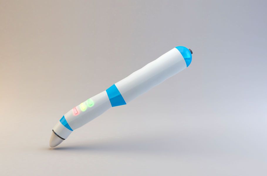
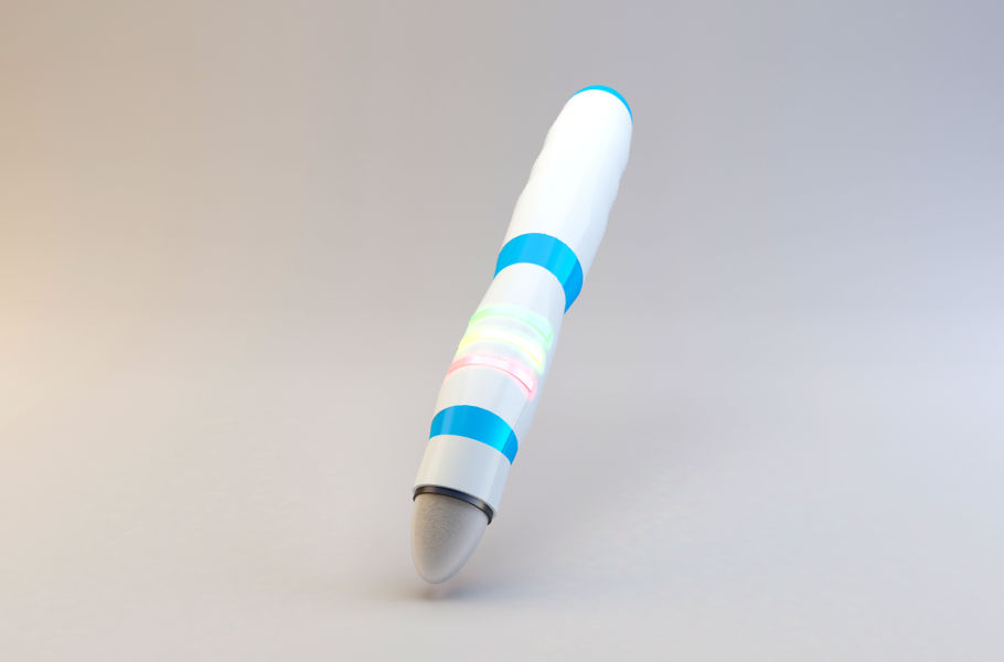
Innovative intraoperative probe for breast cancer detection
The ONCOscanner is an oncological probe for identifying cancerous tissues during surgery, specifically designed for breast cancer procedures.
High precision
The probe’s effectiveness has been confirmed in clinical studies with several hundred patients.
Real-time information
The only device that enables intraoperative assessment of healthy tissue margins.
Usability
Ergonomic design and easy result interpretation tailored to surgeons’ needs.
Reliability
A rigorously tested design crafted from high-quality materials for consistent reliability.
How does ONCOscanner work?
The probe detects differences in electrical properties (such as permittivity and conductivity) between healthy and cancerous tissues. In breast cancer cases, healthy tissue resembles fat tissue in its dielectric properties, while diseased tissue is more similar to muscle.
Does the probe only distinguish between healthy and diseased breast tissue?
The method is effective in cases where there is a significant difference in electrical properties between healthy and diseased tissue. In humans, such cases include breast cancer, atherosclerosis (plaque calcification), or lymph nodes altered by cancer (including sentinel nodes sampled during breast-conserving surgery). Among these applications, the method is particularly effective for breast cancer detection.
Has the probe been tested in clinical trials?
Our probe has undergone clinical testing on several hundred patients. The tests demonstrated its ability to accurately identify cancerous tissues, achieving sensitivity and specificity rates of 93% and 87%, respectively, across 193 samples from 70 patients. This confirms it as a precise, easy-to-use device for intraoperative breast cancer localization.
Why ONCOscanner?
ONCOscanner meets the growing need for devices that can identify cancerous tissue intraoperatively, particularly during breast cancer surgeries.
Breast cancer incidence continues to rise globally, with approximately 1.7 million new cases diagnosed annually. It represents 25% of all cancers among women and is the fifth most common cause of cancer-related deaths, accounting for over 500,000 deaths annually.
A precise breast tissue resection reduces complications. Currently, surgeons rely on visual and tactile methods to assess tissue during surgery. If cancer cells are found in the excised tissue margins, the risk of recurrence increases, often requiring additional surgery.
As early detection improves and breast cancer cases increase, more breast-conserving surgeries are performed. ONCOscanner offers a novel solution by providing accurate tissue differentiation, which supports surgeons in achieving clear margins and reduces the reliance on visual inspection alone.
Team
Kazimierz Orzechowski, PhD
Principal Investigator
Marek Rząca, MD
Oncologic Surgeon
Stanisław Baj
Lead Engineer & Designer
Partners
Regional Specialist Hospital in Wrocław
Research and Development Centre
University of Wrocław
Faculty of Chemistry
Become an ONCOscanner partner!
We are looking for:
- Partners to distribute medical devices for oncological surgery
- Investors interested in supporting our company in the next investment round to bring the probe to market
If interested, please contact us at biuro@oncoscanner.com.